OUR MRI COMMITMENT TO PATIENTS & PHYSICIANS
At Salina Ortho MRI, we value the opportunity to serve our patients day in and day out. We are committed to giving our patients the best experience possible while supporting our physicians that refer and entrust their patients to us everyday. They know we treat each of them as if they were family.
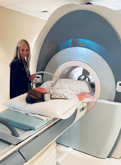
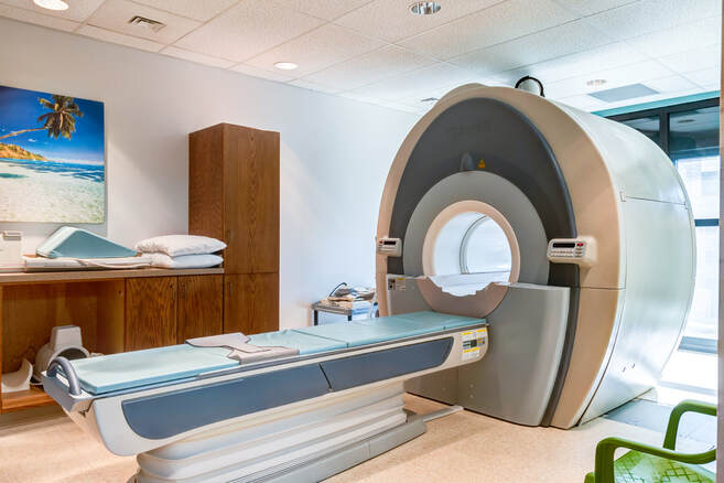
It is important to us that we are transparent with each of our patients and our community. Patients can save up to $1000 or more by having their MRI's performed at Salina Ortho. In some cases we are half the price of what a patient may pay should they go elsewhere. We are able to offer same-week appointments and results within 48 hours. We have a TOSHIBA Vantage 1.5 Tesla Magnet. This MRI scanner has an ultra short bore to help reduce the feeling of claustrophobia.
If your clinic or doctor’s office has a specific MRI protocol they prefer - we will work with them to ensure the exact images and angles needed are retrieved in order to provide a more precise diagnosis.
|
Overview
Magnetic resonance imaging (MRI) is a medical imaging technique that uses a magnetic field and computer-generated radio waves to create detailed images of the organs and tissues in your body. MRI is a noninvasive way for your doctor to examine your organs, tissues and skeletal system. It produces high-resolution images of the inside of the body that help check for abnormalities and diagnose injuries and conditions. MRI is a painless procedure that simply requires that patients remain still while the images are taken. Risks Because MRI uses powerful magnets, the presence of metal in your body can be a safety hazard if attracted to the magnet. Even if not attracted to the magnet, metal objects can distort the MRI image. Before having an MRI, you'll complete a questionnaire that includes whether you have metal or electronic devices in your body. Unless the device you have is certified as MRI safe, you may not be able to have an MRI.
|
If you have tattoos or permanent makeup, ask your doctor whether they might affect your MRI. Some of the darker inks contain metal.
Before you schedule an MRI, tell your doctor if you think you're pregnant. The effects of magnetic fields on fetuses aren't well understood. Your doctor might recommend an alternative exam or postponing the MRI. Also tell your doctor if you're breast-feeding, especially if you're to receive contrast material during the procedure.
It's also important to discuss kidney or liver problems with your doctor and the technologist, because problems with these organs might limit the use of injected contrast agents during your scan.
Preparation
Before an MRI exam, eat normally and continue to take your usual medications, unless otherwise instructed. You will be asked to change into a gown if you have any metal attached to your clothing and to remove things that might affect the magnetic imaging, such as:
Before an MRI exam, eat normally and continue to take your usual medications, unless otherwise instructed. You will be asked to change into a gown if you have any metal attached to your clothing and to remove things that might affect the magnetic imaging, such as:
Tell the staff if you have a pacemaker, cochlear implant, surgical hardware, breast tissue expander. People with heart pacemakers should not have MRIs. Let the staff know if you have claustrophobia, a fear of small confined spaces. You may receive a sedative prior to the procedure to help you relax. You will receive specific preparation instructions when you schedule your appointment. |
Procedure
The MRI machine looks like a long narrow tube that has both ends open. You lie down on a movable table that slides into the opening of the tube. A technologist monitors you from another room. You can talk with the person by microphone.
If you have a fear of enclosed spaces (claustrophobia), you might be given a drug to help you feel sleepy and less anxious. Most people get through the exam without difficulty.
The MRI machine creates a strong magnetic field around you, and radio waves are directed at your body. The procedure is painless. You don't feel the magnetic field or radio waves, and there are no moving parts around you.
During the MRI scan, the internal part of the magnet produces repetitive tapping, thumping and other noises. You might be given earplugs or have music playing to help block the noise.
In some cases, a contrast material, typically gadolinium, will be injected through an intravenous (IV) line into a vein in your hand or arm. The contrast material enhances certain details. Gadolinium rarely causes allergic reactions.
You will lie on a narrow table, and the technician will position your body. Small devices, called coils, may be placed by your body to enhance the images.
Once you are positioned, the technician will step back into the control room. You will be able to communicate with the technician via a microphone at all times. The table will slide into the MRI machine. You will be asked to remain motionless while the images are taken. An MRI can last anywhere from 15 minutes to more than an hour. You must hold still because movement can blur the resulting images.
A doctor specially trained to interpret MRIs (radiologist) will analyze the images from your scan and report the findings to your doctor. Your doctor will discuss important findings and next steps with you.


